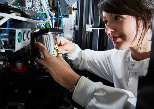
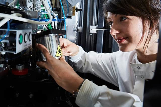
Ultra-powerful microscopes at Leeds University help tackle Alzheimer's


Scientists at the University of Leeds used multi-million pound electron microscopes to make a breakthrough in the understanding of the causes of the illnesses.
The state-of-the-art instruments enabled them to reveal the structure of amyloid, a build-up of abnormal proteins in the body linked to Alzheimer’s, Parkinson’s and Huntingdon’s disease.
Advertisement
Hide AdAdvertisement
Hide AdThe research team said their findings could be used by drug manufacturers to develop cures for amyloid illnesses.


A five-year project using two 10ft cryo-electron microscopes was led by Professors Sheena Radford and Neil Ranson at the university’s Astbury Centre for Structural Molecular Biology.
Prof Radford said: “We’ve used cryo-electron microscopy not only to uncover the shape and structure of amyloid proteins, but also how they grow and intertwine with each other, like the strands in a rope, to form larger assemblies.
“This knowledge is going to be crucial for knowing how to deal with them.”
Advertisement
Hide AdAdvertisement
Hide AdHigh resolution images of the proteins have been published in the journal Nature Communications.
Prof Ranson said: “Until a year or so ago, scientists knew the structure looked more or less like a ladder, but we have now shown it is much more complex than that. We’re now beginning to see how different proteins folded up into different shapes and how those vary with every disease they cause.
“The extra detail we have uncovered means we can start to understand these proteins’ disease-causing abilities.”
The team worked with Professor Bob Griffin at the Massachusetts Institute for Technology, on the study.
Further research is planned which could lead to the involvement of a drug manufacturer, Prof Ranson added.