Julian Norton: Rescue therapy needed for Suki


Nevertheless, Suki was blissfully unaware of her illness. There is often some chit-chat in the waiting room at Skeldale,
“What’s he in for?” or “That’s a big bandage - what’s been happening to you?”
Advertisement
Hide AdAdvertisement
Hide AdIt is often the case that the seriousness of a chemo patient’s illness goes unnoticed amongst the bandaged legs, limps, lumps or stitches of other dogs and cats. Suki mascaraded as a normal dog.
As the weeks passed, however, I became increasingly worried about the size of her abdomen. Suki was getting bigger. It was ironic that, surrounded by the myriad of modern diagnostic techniques, like flow cytometry and immunohistochemistry, I reached for the tape measure, that we used to monitor fat dogs, to check on Suki’s progress.
She was also paler than I would have liked and when I looked at the results of her most recent blood test, it became clear that, as Suki’s girth was increasing, her blood cells were falling. It was time to put the tape measure to one side. Suki needed x-rays, and I needed to take aspirates from both her spleen and her bone marrow, to establish whether the cancer cells had spread to other parts of her body. Since Suki was quite used to injections, the anaesthetic presented no traumas and she was soon asleep. The x-rays showed that the increasing size of her abdomen was due to a grossly enlarged spleen, which I carefully sampled. The bone marrow aspirate was slightly more fiddly, but my microscope slides and little pink sample pots were soon ready to send to the laboratory for detailed analysis.
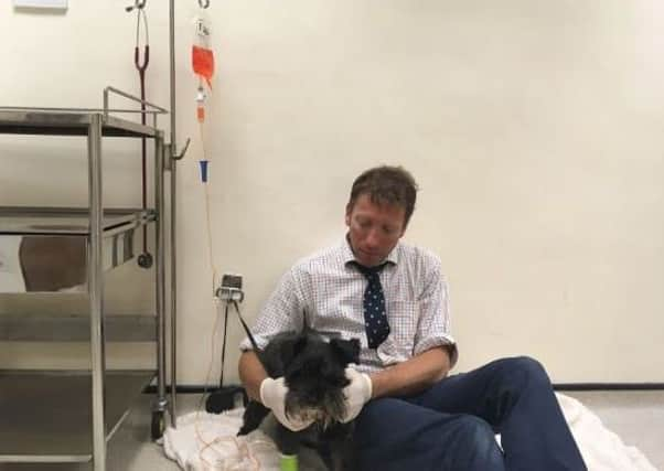
Three days later the lab report arrived. It confirmed what I had feared. Lymphoma cells had invaded Suki’s spleen and also her bone marrow. This explained why she was so pale and so round. A long and detailed conversation followed with Suki’s owners, exploring all the possibilities. In the end, we decided to go ahead with a “rescue” therapy. This involved a complete revamp of the chemotherapy regime. Excitingly for me, the new drug, which needed to be run slowly and carefully into the vein in her front leg through a drip line, was fluorescent orange! As I hooked Suki up to the brightly coloured infusion, heads turned in the prep room - the usual fluid running through a drip is colourless.
Advertisement
Hide AdAdvertisement
Hide AdThen came the best bit. Because the drug is quite toxic, it is important to supervise the animal closely while the infusion is in progress. It would be a disaster if the line were chewed or pulled out. This is what I tell the nurses when they walk past and see me sitting on a fluffy dog bed, cuddling my patient. It is the perfect excuse to sit on the floor and spend 20 minutes with my friend.
The following week, Suki came back in to see me. Her gums were pink and her abdomen was back to normal size. I didn’t need my tape measure to see that she was plunged firmly back into remission.
Julian’s book, Horses Heifers & Hairy Pigs: The Life of a Yorkshire Vet, is on sale now.